Welcome back to The Sydney HEMS Debrief, now on episode 14!
Today, we are joined by Retrieval and Anaesthetic Staff Specialist Dr Jess Devlin, who talks us through one of her most challenging and rewarding cases. Using the case as a springboard, we go on to discuss some of the nuances around, and approaches towards, multi-casualty scenes and consider how to make sense of an overwhelming situation and perform effectively within it.
This episode is certainly a must listen for any clinician who may be faced with a multi-casualty, multi-agency situation, and who wants to function effectively and safely with in.
Welcome back to The Sydney HEMS Debrief, now on episode 13.
Today we are talking about the PreCare trial, currently underway here in Sydney. This feasibility trial – a first for Sydney – concerns placing patients suffering out of hospital cardiac arrest onto potentially life-saving extracorporeal membrane oxygenation (ECMO) therapy in the field, before transporting them to definitive care.
To discuss more, we are joined again by Dr Nat Kruitt, Staff Specialist here at Sydney HEMS and Specialist in Cardiac Anaesthesia and ECMO, as well as Alex Peters, one of the Sydney HEMS Critical Care Paramedics. We will discuss how and why the trial came about, the practical application and workflow considerations of placing an eligible patient onto pre-hospital ECMO, as well as some future considerations for this innovative therapy.
This episode is a must listen for other pre-hospital providers in the greater Sydney basin who may come into contact with the ECMO team, as well as those interested in eCPR and ECMO more broadly.
I hope you enjoy it!
Posted inGeneral PH&RM, Podcasts|Taggedpodcast|Comments Off on HEMS Debrief #13 – The PreCare (ECPR) Trial: Prehospital ECMO in Sydney, Australia
Obtaining access in shocked trauma patients can be notoriously difficult due to circulatory collapse. Those who are shocked, shut down with limited or no other options for peripheral access require central access. The cohort of patients that require this intervention in the pre-hospital setting are likely to be the most critically unwell patients we encounter.
In trauma, access to the internal jugular vein can be difficult due to cervical collars and concomitant airway interventions. Similarly pelvic binders and the potential for sub-diaphragmatic vascular injury can preclude the use of femoral access. This is why the blind subclavian approach is favoured; in experienced hands it is the most anatomically consistent approach.
The evidence for improved safety and quality with the use of ultrasound for CVC implementation is well established[i]. In fact, it’s use is so ubiquitous that the newer generation of emergency physicians are largely inexperienced in the insertion of central lines as a blind technique. This rather disconcertingly means then that the sickest patients we encounter may require a procedure that a significant number of doctors may have had very limited experience with or exposure to during their training. How can this situation be rectified or has the use of ultrasound rendered this blind subclavian “trauma line” a thing of the past?
Subclavian CVC insertion using ultrasound can be tricky due to the difficulty in visualising the vein due to the overlying clavicle. The axillary vein approach/distal subclavian can allow for ultrasound use but it represents a smaller target which may preclude it from being considered in the pre-hospital sphere where environmental factors often mean the set-up is less than ideal, not to mention the increased difficulty in a hypovolaemic shocked patient.
One proposed alternative is using the supraclavicular approach to subclavian vein cannulation. The supraclavicular approach is not a new technique, but it is underutilised. In fact, it was first described by Yofa in 1965 as an alternative to the infraclavicular approach for SCV cannulation[ii]. This approach is a bit of a misnomer as in reality it involves cannulation of the brachiocephalic vein (BCV) origin. The right BCV origin is preferred as it is more superficial, larger and straighter. Its use is perhaps most widely studied in the paediatric population, where this approach is found to have a high success rate and a low procedural complication rate[iii],[iv].
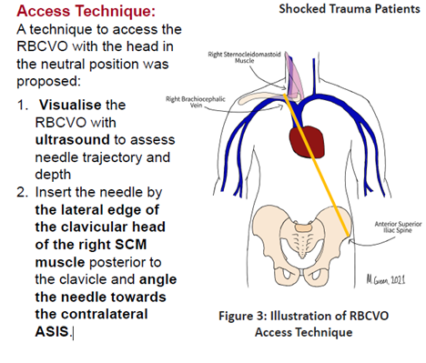
Preliminary studies including a biometric analysis of CT scans and prospective ultrasound study suggest that right BCV origin access is feasible in shocked trauma patients and the RBCV does not collapse in severe shock[v]. The right BCV is preferred as it is more superficial and straight compared to the left BCV which was observed to be deeper and has a more tortuous course[vi]. One approach to obtaining BCV origin access (fig.1) suggests using ultrasound to determine needle trajectory and depth and then proceeding without ultrasound guidance. This would therefore remove some of the limitations (perceived or real) around time delays associated with using ultrasound for central access as there would be no need for probe covers and positioning of the portable ultrasound (which often needs the help of a second operator, a significant limitation in a small prehospital medical team).
A small single centre study conducted in an Australian tertiary trauma centre on shocked trauma patients presenting to ED showed that right BCV access is feasible and had a higher success rate for 1st attempt access than the subclavian vein (63 vs 48%)[iv]. Some limitations were noted however in visualisation of the vein with ultrasound in obese patients and those with subcutaneous emphysema.
Perhaps more importantly this technique may serve to bridge the gap between the newer generation of critical care physicians who may be more reluctant to adopt a completely landmark base/blind approach to central access. At the very least, knowledge of it as an option in the armamentarium of retrieval and critical care providers is important. It is currently the subject of future research and publication in the emergency department setting. Its use in the prehospital environment is not well known or studied and may be an interesting area for future research.
[i] Leibowitz A, Oren-Grinberg A, Matyal R. Ultrasound Guidance for Central Venous Access: Current Evidence and Clinical Recommendations. Journal of Intensive Care Medicine. 2020;35(3):303-321. doi:10.1177/0885066619868164
[ii] Yofa D. Supraclavicular subclavian venepuncture and catheterisation. The Lancet. 1965;286:614–7
[iii] Breschan C, Platzer M, Jost R et al. Consecutive, prospective case series of a new method for ultrasound-guided supraclavicular approach to the brachiocephalic vein in children. Br. J. Anaesth. 2011; 106: 732–737.
[iv] Breschan C, Graf G, Jost R et al. A retrospective analysis of the clinical effectiveness of supraclavicular, ultrasound-guided brachiocephalic vein cannulations in preterm infants. Anesthesiology 2018; 128: 38–43.
[v] M. Green et al. Right Brachiocephalic Vein Origin Access for the Resuscitation of Shocked Adult Trauma Patients. Poster presented at Australia, New Zealand Trauma Society Conference, Melbourne, VIC, Australia. October 2023.
[vi] Xia R, Sun X, Bai X et al. Efficacy and safety of ultrasound-guided cannulation via the right brachiocephalic vein in adult patients. Medicine 2018; 97: e13661.
Sydney HEMS acknowledges the Australian Aboriginal and Torres Strait Islander peoples as the first inhabitants of the nation and the traditional custodians of the lands where we live, learn and work.