A significant number of our critical care patients are dependent on vasoactive drugs, so it is worth reviewing the process for managing these infusions during inter-hospital transfers.
The priming volume of the distal lumen of a central line is 0.44mls. Therefore an infusion running at 10mls / hour could take approximately 3 minutes to reach the patient if started de novo. The half-life of inotropes such as adrenaline and noradrenaline is short, approximately 1 minute. This leaves the patient vulnerable to a period of hypotension. Corrective bolusing of vasoactive drugs is not recommended as it can lead to large, and potentially detrimental, swings in blood pressure. Therefore a smooth transition between syringe pumps is recommended to maintain haemodynamic stability.
There are a number of different methods for double pumping but one suggested protocol is as follows:
Leave infusion number 1 running at its current rate (100%)
Commence infusion number 2 at 50% of the rate of infusion number 1
Wait for a small kick in blood pressure to indicate that the second infusion is reaching the patient. This can be up to but should not exceed SBP 20mmHg
Immediately increase infusion number 2 to 100% (the current rate of infusion 1) and reduce infusion number 1 back to 50%.
Reduce infusion 1 back to zero incrementally over a few minutes ensuring that BP doesn’t drop.
Tips and tricks
Avoid the use of a 3-way tap if you have enough ports to manage without. Three-way taps increase the complexity of the process and the likelihood of an error. No matter how intelligent you think you are, 3-way taps have a habit of embarrassing you. If you really must use a 3-way tap make sure both taps are “on” to the patient during the process and then make sure the correct tap is left “on” at the end.
Dedicate a member of the HEMS team to the double pumping process and avoid being interrupted during it.
Use a dedicated line for vasoactive drugs to prevent inadvertent bolusing
Label the line and the syringe driver clearly to prevent confusing the vasoactive drug with sedation (which can be, and often is, bolused)
If you are about to embark on a long transfer you may want to replace a single strength vasoactive drug with double strength. Take this into account when double pumping. If infusion 2 is double strength it will need to be started at 25% of infusion 1 and then increased to 50% once the BP kick is seen.
Be patient. Depending on the infusion rate and the dead space in the catheter lumen, the process can take several minutes. It is worth taking the time to do it smoothly rather than trying to speed it up by increasing infusion rates or bolusing.
The following learning points are collated from Regional and Rural hospitals of NSW, Australia, represented here by a single fictional institution – Umbara Base Hospital. Cases are amalgamated and anonymised (including alteration of patient demographics) such that similarity to real patients is coincidental.
Below are some high-yield learning points collated from the Umbara Hospital trauma case review meeting.
Beware distracting injury
Patients with blunt trauma with significant orthopaedic injury may have other significant other injuries that are difficult to assess on history and examination and easy to miss.
Anchoring bias may also occur, particularly in inter-hospital transfers. These are higher risk patients for missed/delayed diagnosis. Reassess the patient from the start.
Rib injuries in the elderly
Elderly patients (>65) who sustain blunt chest trauma with rib fractures have twice the mortality and thoracic morbidity of younger patients with similar injuries. For each additional rib fracture in the elderly, mortality increases by 19% and the risk of pneumonia by 27%.
CXR is inaccurate in diagnosing the presence and number of rib fractures – hence we should have a low threshold for CT to further assess. Current best practice is the ChIP protocol.
Angioembolisation
Angioembolisation in renal trauma is effective in selected patients.
Trauma CT: Blue arrow is contrast ‘blush’=active bleeding point. Red arrow is perinephric haematoma.
Angioembolisation. Red arrow=coils in arcuate artery of kidney which has stopped the bleeding.
When transferring these patients from smaller hospitals to larger centres, consider whether initial destination should be the ED for rapid re-assessment prior to entering IR suite.
Stab Heart
Anterior and posterior ‘cardiac box’
Penetrating trauma to the ‘cardiac box’ may result in cardiac injury and pericardial effusion leading to tamponade. A permissive hypotension strategy is followed where practical.
The following learning points are collated from Regional and Rural hospitals of NSW, Australia, represented here by a single fictional institution – Umbara Base Hospital. Cases are amalgamated and anonymised (including alteration of patient demographics) such that similarity to real patients is coincidental.
Below are some high-yield learning points collated from the Umbara Hospital trauma case review meeting.
Chest drains
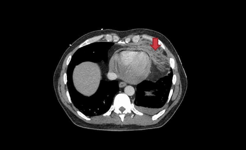
Always check the chest drain with a CXR – particularly check for the drain’s position and for complications such as kinking, as can be seen in this left sided CXR.
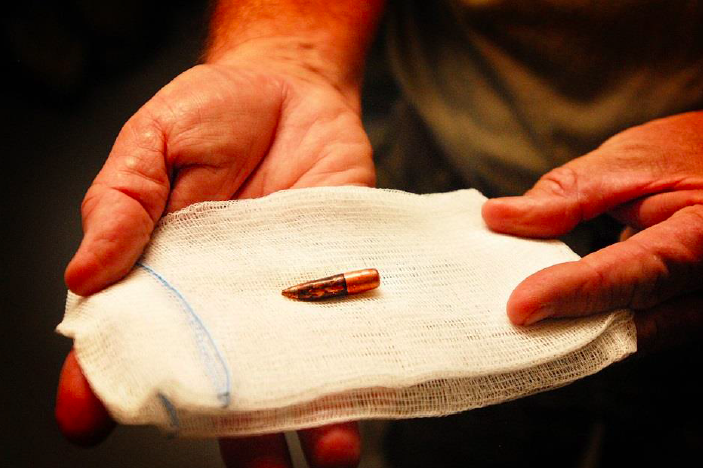
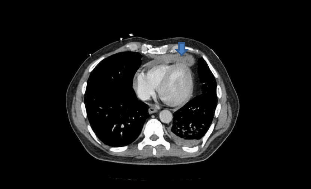
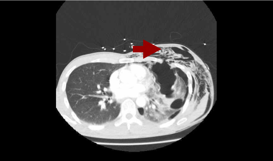
Pneumothorax is often associated with subcutaneous emphysema (free air in the tissues under the skin). It feels like bubble-wrap and looks like this on CT. Don’t press too hard though – it’s likely there are rib fractures underlying the air and they are extremely painful!
Elderly trauma
Trauma in older patients is increasing with our ageing population here in Umbara – ever improving management of chronic health conditions means people are living longer. It is difficult to predict mortality in elderly trauma and hence some scoring systems exist.
These scoring systems may help guide discussions with patients and their families in the future.
Non-accidental Injury in Children
Data from the UK Trauma Audit and Research Network (TARN) showed that 2.5% of children in their database had suspected child abuse underlying their injuries. 97.7% of these children were aged <5yrs; 76.3% were aged <1yr. Injury severity score (ISS) was also greater in patients with suspected child abuse: they were 1.7x as likely to have an ISS score >15.
IDENTIFY risk factors through history, examination, observation.
LISTEN and watch parent-child interaction
CONSIDER the possibility
DON’T DISMISS non-accidental injury as a possibility due to lack of physical findings
DOCUMENT carefully, clearly and contemporaneously
PREVENT by linking with services
KNOW your legal requirements for reporting
Risk Factors for NAI
Although non-accidental injury can occur in the absence of these factors, there are several factors which have associations with non-accidental injury.
In the child:
Chronic illness, disability or developmental problem
Prematurity
Age of child
“Difficult” behaviour
Parent/Family:
Unwanted pregnancy
Young parents
Single parent family
Relationship problems
Exposure to drug and alcohol abuse and/or family violence
Low socioeconomic status
Social isolation
Physical or mental illness in a parent
Other concerning features:
Poor hygiene
Dirty clothes
Missing a lot of school
Previous contact with FACS / CPU
Clinical/Attendance features:
Delay in presentation
Injury not explained by story
Inconsistent with developmental ability (know developmental milestones! Here’s a quick reminder)
Inconsistencies in history and changes over time
Unexplained or unwitnessed fall with neglect
Previous suspicious injuries
Unusual parent – child interaction
Failure To Thrive (FTT)
Resuscitation efforts caused injuries
Patterned bruise/burns; certain distribution
Spiral/transverse long bone fractures, particularly in non-mobile children
When Umbara Base Hospital’s own Dr Tallie fell from her horse earlier this month and ended up being treated in her own Emergency Department, she was in a unique position in being able to provide constructive feedback around her own care with a full understanding of the processes of the hospital.
This month we invited her to share her thoughts at the trauma case review meeting and were delighted to learn the following.
Prehospital methoxyflurane is an excellent analgesic and she was very grateful for it
She found the experience had increased her trust: she was happy to put her life in hands of the staff of Umbara Base Hospital and has a renewed appreciation for them all as a result
She particularly noted that nurses are awesome (both during her ED and ICU stays): she added, “it’s the ‘little things’ that make all the difference”.
Having experienced both, she found regional block much better in the pain management of her rib fractures than drugs.
Overall she was dismayed to realise just how long bones take to heal.
Huge thanks to Dr Tallie for her insights – we wish her well on her recovery.
Drs Natalie May, Geoff Healy, and Cliff Reid discuss missions in which the prehospital medical team is diverted to a hospital because their patient has been moved from scene to a (non-major) hospital.
While one might expect these missions to be easier from the point of view of having the hospital environment and resources to hand, they can in fact be more complicated and take longer than typical prehospital (primary) and interhospital (secondary) missions.
There are a number of reasons for this:
Timing – the retrieval team (RT) usually arrives shortly after the patient, disrupting the hospital team (HT)’s initial assessment and management
Clash of goals – RT focuses on rapid management of essentials and extrication of the patient to a trauma centre. HT often focused on more traditional ATLS management
Assumptions – RT uniform and appearance may make the HT assume the RT are the paramedics who brought the patient in, and dismiss their attempts to offer advanced interventions and leadership
Crowd control – large number of good people keen to do things, can paradoxically make interventions much longer to perform compared with a small well rehearsed RT working to mutually understood operating procedures.
Nat, Geoff and Cliff discuss these challenges and how to tackle them, including how to introduce yourself, what language to use, how to integrate yourself into the team and offer support and if necessary leadership, the importance of a collaborative approach, and the critical contribution of the RT paramedic in making all this happen smoothly.
This second post from the Sydney HEMS ECMO education days summarises a great session by Retrieval Doctor and all-round legend Natalie Kruit on Troubleshooting in ECMO Retrieval.
What’s the role of the Retrieval Doctor?
As mentioned in Part One, the retrieval doctor is essentially there to look after the patient as a whole, while the separate ECMO team will oversee the function of the ECMO pump.
The first thing to know as a retrieval doctor is that the pump is primed with crystalloid – there is approx. 1L in circuit in total – so we should expect patients to be a bit unstable after transition onto pump!
VV ECMO
There is an access line in or near vena cava – this draws blood into the pump. This is the negative pressure part of the circuit.
Blood drains into centrifugal pump, is oxygenated and is returned to the right atrium.
VV ECMO inclusion criteria
The Murray score is used for this: for a score >3 – consider referral to ECMO centre
Murray score: VV ECMO inclusion criteria
Pathologies that will do well: asthma, ARDS. The longer they’ve been on a ventilator, the less likely they are to do well on ECMO.
What can you as the retrieval doctor do?
Optimise
Sedate and paralyse
Find best available vent
Recruit and PEEP
Diurese
HRCT
Right radial arterial line for sampling etc
Troubleshooting 1: Access Insufficiency
This occurs when the vein the cannula is sitting in is collapsing around the cannula.
Treatments: drop the revs, give volume, remove obstruction, consider and exclude tamponade
Ventilation strategies on VV ECMO
Once on VV ECMO – target lung protective ventilation: rest the lungs, minimise alveolar strain
Reduce FiO2 (target around 0.6 if possible: O2 toxicity, diffusion atelectasis from reabsorption), reduce the RR, increase PEEP (prevent alveolar closing and opening – maintain alveolar opening), smaller TVs (4mls/kg or even lower) (letting the ECMO circuit do the oxygenation)
Troubleshooting 2: Bleeding on ECMO
Caused by
Fibrinolysis
Circuit driven fibrinolysis
Mucosal bleeding initially
Strong correlation with mortality
Heparin resistance
Targets: Platelets >50, Fibrinogen >1.0, INR <2.0, APTT 1.5x normal
If bleeding: TXA, FFP, platelets (prepare to do an ECMO circuit change as may clot) – don’t give protamine (you will get clots and that is maximum badness!
Troubleshooting 3: Hypoxia
When SpO2 increasing on ECMO machine, the patient becoming hypoxic – this suggests recirculation (blood not getting to the patient)
Shunting on VV ECMO
The circuit may not be able to capture full cardiac output, so some deoxygenated blood is circulating. Preferentially this seems to go to the brain and heart. This can happen in someone whose circulation is hyperdynamic (eg sepsis, morbid obesity). The drainage cannula is a good determinant of flow (a bigger drainage cannula gives better flows).
Cardiac output >ECMO flow (if you set flow at 5L/min and the patient is hyperdynamic at 10L/min, only half blood vol will be going through circuit and being oxygenated)
Treatment:
Increase ECMO flow (but you risk suckdown)
High flow configuration – consider a second access cannula (can add third access line in neck if in fem/fem configuration)
Increase O2 carrying capacity: transfuse if Hb is low
Check oxygenator (may be cause due to clots) – measure pre and post oxygenator O2. Look for delta P <50
Check for recirculation (see below)
Check ventilator settings
Troubleshooting 4: Recirculation
This occurs when the access line and drainage line are sitting too close together so the flow is bypassing patient altogether! Or if there is increased intrathoracic/intracardiac pressure (e.g. tamponade)
You may see a low arterial SaO2 and high access cannula SaO2.
Treatment:
CXR to check pipe position
Could withdraw appropriately to increase distance between drainage and return lines
Check for tamponade
VA ECMO
There are two different cannulation options for VA ECMO.
Central – directly cannulated via sternotomy
Peripheral – femoral vessels (blood from the femoral vein flows out to pump then back into femoral artery and pumped UP – this is non-physiological! Blood will be flowing against the normal flow direction – towards heart. This configuration needs a backflow cannula to ensure distal limb perfusion)
You’ll get some mixing of the deoxygenated blood from the ventricle with the oxygenated blood from the ECMO circuit – the more contractile the ventricle, the more distal the mixing point. There’s a nice interactive critical care case here that covers some of this.
If the lungs are not working and the heart is starting to recover and therefore has some recovering ejection fraction, the heart will preferentially eject to coronary and cerebral circulations (so check the ECG for signs of cardiac ischaemia) – it’s also helpful to have a SpO2 probe on right hand to see differential hypoxia early.
— Medlife Crisis (DEFUNCT) (@MedCrisis) June 2, 2016
In this scenario the heart is pumping (so there is a native cardiac output) and that is picking up and distributing deoxygenated blood that we can’t capture with ECMO flow. You may see demarcation of skin tone change across the trunk and/or the ECG may show ischaemic changes. There’s a nice diagram here which comes from this open access paper in Circulation (clicking the link will download the PDF).
Treatment:
try to treat the lungs (eg bronchoscopy, increase the PEEP, add a venous catheter so some oxygenated blood is going to right heart)
Patient selection for VA ECMO
Patients unable to wean from bypass (post cardiotomy)
Awaiting heart transplant
RV failure
Overdose
AMI with multi-organ failure
Cardiomyopathy/myocarditis
When severe end organ damage has occurred (e.g. AST/ALT very high, lactate >10 and not clearing) we have missed the boat!
Ventilation strategies on VA ECMO
We are aspiring to the least damaging lung ventilation
Maintain PEEP (aeration)
Measure SpO2 and PaO2 right arm
Minimise RV afterload and strain – avoid hypercapnia, hypoxia and complete lung collapse
Haemodynamic Management
Discuss haemodynamic goals with ECMO specialist
Inotropes to maintain ejection fraction
Maintain perfusion pressure for end organ perfusion
Promote forward flow: maintain MAP 65mmHg ideally (less than 90mmHg)
Troubleshooting 2: Loss of pulsatility
Causes:
Tamponade
Myocardial depression
LV failure
Access insufficiency will be the first sign – blood is stuck in lungs not getting to circuit
You shouldn’t need to give a fluid bolus – so if doing this, consider LV failure is evolving (dilating LV with inability to pump out)
Offloading the LV – may need to decompress by creating a vent between L and circuit, R heart – there’s potential to use impella to suck blood out directly
Retrieval Final Checks Before Departure
Adequate oxygen and power supply
Confirm haemodynamic goals with ECMO specialist
All taps have stopcocks on
Access line dark, return line bright
Circuit flows stable
Lines secured, away from snag hazards
Patient sedated and paralysed
If you work for Sydney HEMS, take a look at the new ECMO class available through the virtual learning environment.
A/Prof Paul Forrest: VA ECMO in Sepsis
A/Prof Forrest talked a little bit about VA ECMO in sepsis.
The key to the septic shock definition is MAP 60mmHg (or inotropes required to keep it here).
Most adults dying from sepsis do so in a high output state (increased cardiac output with decreased systemic vascular resistance): this paper from 1984 looked at 20 patients with severe septic shock – 50% had moderate to severe decrease in left ventricular function. Notably they found the left ventricular ejection fraction had returned to normal within ten days in those who survived.
This leads us to consider whether low ejection fraction might be a good thing in patients with septic shock – this review by Vieillard-Baron et al from 2001 seems to support the hypothesis.
It’s possible, then, that the relationship between survival and non-survival depends on systemic vascular resistance in the context of vasoplegia – this allows for left ventricular ejection fraction to be maintained against lower resistance
There’s some evidence we might consider in paediatric patients:
They looked at 45 patients, of whom 21 survival (47%). In terms of central vs peripheral VA ECMO, survival proportions were 73% vs 44% and no survivors severely disabled. The authors felt that their data suggested that central configuration might provide a survival advantage.
The same team published a further paper in 2011: in this paper they studied 23 patients, 17 of whom survived to discharge (74%): they noted that a high lactate predicted worse survival.
For adults the data is lacking; literature only describes around 200 adults on VA ECMO when case series are combined and there are considerable different baseline characteristics between patient groups.
There is also the possibility (in adult patients) of using V-AV ECMO: this is when a second return line is sited in the internal jugular (or similar) with the aim of providing oxygenated blood to the right heart. The femoral artery is part of a higher pressure circuit than the internal jugular line and it can be tricky to regulate flow between the two lines. As yet there is little data on this: this case series by Yeo published in Critical Care in 2016 reports a 50% survival with this technique for adults with severe ARDS and septic shock.
Indications for VA ECMO in refractory septic shock
For paediatric patients – central VA ECMO seems the best option
For adult patients:
In the presence of severe ARDS: V-AV ECMO
In the absence of severe ARDS: VA ECMO
VA ECMO in septic shock
In summary, VA ECMO in septic shock seems to be good for:
Kids with or without CA
Adults with low cardiac output/high systemic vascular resistance state
Most of the following post is taken from the recent Sydney HEMS ECMO Education day with some taken from an earlier ECMO education day and amalgamated into a huge amount of notes.
ECMO in New South Wales
Associate Professor Paul Forrest, a Cardiothoracic Anaesthetist and ECMO Specialist from RPA Hospital kicked off the day with an overview of the evolution and current state of ECMO services in NSW. He has a full uploaded presentation here you might be interested in or you can watch the full video below.
ECMO team: Surgeon/anaesthetist or intensivist, medical perfusionist +/- technician
Retrieval team: Doctor and paramedic/nurse
Where do patients go for ECMO?
Patients are routed to St Vincent or RPA as per the Medical Retrieval Unit who oversees the mission.
When did the service start?
The initial service commenced prior to the H1N1 outbreak – this paper in JAMA Oct 12 2009 describes how the service was utilised during the outbreak. 68 patients were treated with ECMO for a median of 10 days (IQR 7-15 days) with 48 surviving to ICU discharge (32 to hospital discharge, 16 still inpatients) at the time of publication.
How is ECMO undertaken during retrieval?
The Cardiohelp pump is used with a customised footplate. When an ECMO team is tasked for a retrieval mission, the team will come with four custom packed retrieval packs of their own.
58% of ECMO transfers are transported by road, 27% by fixed wing and 15% by helicopter.
How successful is ECMO in NSW?
Feb 2016 figures: 68% survival to discharge overall, 72% in resp subgroup and 60% for cardiac subgroup
The St Vincent’s team, headed up by the wonderful Sean Scott, joined us for an ECMO themed sim. Learning was centred around an interactive simulation, the retrieval of a 55 year old patient from a smaller hospital in conjunction with the ECMO team.
Our role in these missions as the retrieval team: to take care of the patient, while the ECMO team takes care of the ECMO machine and pipes (“it’s not about the plumbing”). The full talk is shown in the video below.
ECMO Basics
The Two Types of ECMO
ECMO is basically a heart and a lung – a pump and an oxygenator. Where you plug in the pipes determines the sort of support you (the patient) get(s).
VV – oxygenation, no cardiovascular support
VA – also generates a blood pressure, essentially a cardiopulmonary bypass machine
We should consider ECMO as a bridge; generally it is used as a bridge to recovery but sometimes to a decision (further resuscitation? Transplant?) or definitive intervention (treatment of cause of cardiac arrest).
Why (when) VV?
This is effectively lung bypass, used when the “lungs don’t work” – there are many causes, for example:
ARDS
Airway problems (can’t be intubated eg airway injury)
Bad asthma
Needs to be a potentially reversible cause
Why (when) VA?
This is effectively heart and lung bypass, used for support in circulatory failure – cardiogenic shock is the most common/obvious reason (although the underlying cause may be MI, overdose etc.)
Trauma? – has been used in hypotensive trauma patients
Sepsis? – jury is out on whether this is a good idea (how reversible are the processes involved?)!
How does it work?
Most common is femoral/femoral VV cannulation
Fem/fem – two cannulae in IVC, one draining and one returning – can’t use two multistage cannulae (recirculation – when blood doesn’t actually flow through the circulation – is very high)
Fem/jug placement avoids this
What about in Cardiac Arrest?
Now features in the 2015 ALS guidelines under “consider” (extracorporeal CPR)
2CHEER (the study into LUCAS plus ECMO for out-of-hospital VF arrest) is ongoing in Sydney
However, it doesn’t “consider” all the logistical work involved in getting people onto ECPR
ECPR means we should add a few new links to the chain of survival – mechanical CPR on scene, ECMO in ED, straight to cath lab
Common patient scenario – refractory VF, PEA or asystole but after ECMO perfuses the coronaries, ROSC may occur quickly.
ECMO remains on after ROSC (the flow may be dropped a little) – the patient is usually in cardiogenic shock so needs the ongoing support.
How has ECPR been achieved?
Pit crew concept – everyone has defined roles, printed on role cards which they wear
This video shows how this is trained for through simulation.
LUCAS is used as a bridge to ECMO, ECMO is used as a bridge to intervention.
The target of ECPR is maintenance of cerebral perfusion while reversible causes are addressed.
Prehospital Training for 2CHEER was key, with a strong focus on changing the dynamics of arrest calls.
Put LUCAS on early, decision for eligibility early, transport early (with definitive airway if possible)
BatPhone activation to the ECMO centre is essential for team preparation
Clear role development and allocation with role cards (A5)
This training is not particularly frequent – one a month or so – which is helpful for refreshing and framing behaviour.
Should everyone get ECPR?!
No! There are STOP criteria – although in practical terms it is advised that processes continue until there is a reason to stop
>70yrs
Unwitnessed
>10mins without BLS
>60mins since collapse
Initial rhythm asystole
Organ failure or malignancy
No prospect of reversal
How do you echo with all this going on?
Transoesophageal Echo is used to guide cannula placement
There is a strict 90mins cutoff – if the patient has not been (ECMO) cannulated by 90mins from arrest time, efforts should stop (in reality this means the patients need to arrive in ED at 60mins from arrest time). No cases in the Sydney registry have not achieved this as yet!
Anticoagulation/Antiplatelets and Clots
5,000 units of heparin are given when the cannnulae go in. An extra (approx.) 7,000 units are given with the pump (which is primed with 10,000 units, there is some loss during connection).
The venous line is connected first due to risk of clots – they would then be sucked into unit, not into brain (as would occur if arterial line placed first).
What if the cause of arrest is a SAH?
VF arrests can occur with SAH – 15-20% of patients on ECMO end up with ICH (pre or resultant), CT brain is deferred until after definitive cardiac intervention, which may be >24h.
What about for massive PE?
The team aims to get the patient onto ECMO on then thrombolyse – there is a potential for catheter directed lysis. Half dose used thrombolysis has been used as per MOPETT until now but radiology support for catheter directed is now available.
Who is suitable for eCPR after out-of-hospital arrest?
Patients <65
Witnessed VF arrest
Bystander CPR
In-hospital, the team might be more lenient with rhythm (but would still need to know there’s a reversible cause)
Who is not suitable?
Elderly
Comorbidities e.g. liver, renal failure, severe respiratory disease
Advanced malignancies
Advanced care directive
See STOP criteria
What are the common complications?
Usually complications occurring immediately are related to cannulation – bleeding, haematoma, kinked wires, femoral nerve damage, arterial injury.
In standard elective VA ECMO, clinicians would put in a backflow cannula from arterial cannula to perfuse the distal limb. Smaller cannulae are used in ECPR to facilitate distal perfusion; after angiography, a backflow cannula is sited.
Longer term complications include: bleeding/clotting, infection plus the complications of a long ICU stay
Making ECPR happen in the Emergency Department
What happens in the Precannulation phase?
(this is essentially a checklist for the ED Consultant)
High quality CPR
LUCAS
Airway secure
EtCO2
Expose and shave groins
IV access
Cath lab aware
What happens in the Cannulation phase?
Can stop LUCAS but priority is excellent ongoing CPR
Defibrillation is not necessary during the cannulation phase
What happens Post-Cannulation?
Stop LUCAS
Defibrillate (three shocks max)
Get ejection fraction from echo
Expedite transfer to cath lab
Cannulation issues
Immediate term: difficulty getting access, wrong vessels (VV in arrest, AA), haematoma formation – surgical backup (for cutdown) and TOE confirmation can be helpful
Checklists are available and helpful e.g. suck down (insufficient volume to fill venous drainage cannula) – see more info in part two
What’s the Evidence for all this?
It’s not awesome as yet – there are not many RCTs.
Sydney HEMS acknowledges the Australian Aboriginal and Torres Strait Islander peoples as the first inhabitants of the nation and the traditional custodians of the lands where we live, learn and work.