Clinical Governance Day – Wednesday 16th May

Posted in General PH&RM | Leave a comment

AiR – Learning from the Airway Registry [January 2018]

Intubations this month:          27

Airway Registry learning points reflect the challenges described and wisdom shared by Sydney HEMS personnel and guests at the Clinical Governance Airway Registry presentation for January 2018. Cases are discussed non-contemporaneously, anonymised and amalgamated over a time period to draw together unifying take-home messages. Details of specific cases are removed and/or changed, such that any similarity to real-life patients or scenarios is coincidental.

All CMAC videos are shared under a Creative Commons Licence: Attribution 2.0 Generic. Please familiarise yourself with the terms of the licence before reusing our videos.

To view these videos, you will need this password: AiRblogVideos

Focus on: Beach

Some of our work takes us to the beach, particularly during the summer months, although the NSW climate is such that beaches are frequented by residents and tourists year-round.

This month we talked about the specific challenges inherent in undertaking our prehospital work in a beach setting.

The Location

  • The downwash of the helicopters used in our service can cause issues with sand blasting the patient, caregivers, and bystanders on landing and take off.
  • Beaches are very public spaces so we may experience reticence from our flight crew towards shut down of the helicopter in beach location (rather than hot offload etc)
  • Crowd control can be an issue, particularly if we are the first asset on scene. We can expect bystanders and onlookers aplenty encroaching on ‘our space’ and this adds to the mental load of the team
  • The inherent instability of sandy surfaces creates challenges for moving and positioning the patient & the patient’s airway
  • Sand is also very ready to swallow our equipment; we should be cautious around placing items on the sand as we may never see them again! This is particularly true of smaller items like syringes or the thoracostomy kit but we should be mindful of the additional challenges of equipment maintenance in these environments
  • For patients who have been immersed, even if they are out of water by the time of our arrival, we might experience airway flooding with sea water or even pulmonary oedema at laryngoscopy

Solutions from the Sydney HEMS Hive Mind

  • Consider the possibility of winching to scene – though there may still be significant downwash generated
  • Clearing the beach whenever possible and using additional resources (Police, road Ambulance crews) to do this
  • Moving the patient to a solid surface or ideally to an ambulance stretcher for RSI
  • Laying a surface covering over the sand to create a treatment area for kit dump. Examples we came up with that might be available to us included
    • a blanket or towel (most road crews have these)
    • a beach towel
    • thermal shock blanket (AKA the “space blanket”) weighted at the corners
  • Anticipate the need for suction and have two sources of suction (ideally) ready

Other Airway soundbites this month

  • Ability to confirm tracheal placement of endotracheal tube can hampered by time for waveform ETCO2 to ‘warm up’ on some monitors if not already warmed up-> remember the EMMA capnometer is widely available from NSW Ambulance crews as well as being carried in our blue prehospital pack.
  • Intubating the acidotic patient – apnoea feeds hypercapnoea and acidosis – hyperventilation is needed to both fight the patients own CO2and that generated by exogenous bicarb administration. Any ventilations during RSI should be done in a way that limits gastric insufflation – we suggest 2 person bag-mask ventilation, limiting inflation pressures and use of a guedel (oropharyngeal) airway.
  • Inadequate paralysis at RSI – note our new RSI operating procedure recommends a dose of 2mg/kg Rocuronium. This gives an average paralysis time of 2 hours from onset.
  • Our training and clinical governance processes for RSI include frequent currencies for doctors and paramedics in our service to maintain efficient team working and safety. Only paramedics and doctors within this clinical governance framework can perform laryngoscopy within our service.
Posted in Airway, Airway Registry, Tips | Tagged , , , , | Leave a comment

Joint HEMS/NETS Education Day: Weds 2nd May 2018

We have been collaborating hard to put together the programme of the next education day with our colleagues across the airfield at NETS. All NSW Health employees are welcome to join us for a day of paediatric and neonatal case reviews, cutting-edge and evidence-based medicine, simulation and workshops. See you there!


Posted in General PH&RM | Tagged , , , | Leave a comment

Clinical Governance Day – Wednesday 18th April

Posted in General PH&RM | Leave a comment

Getting ready for Winter

alpine08 070 copy

Although Sydney is currently enjoying an extended summer, last week’s education day had a frosty feel to it as we were treated to two presentations on the perils of cold weather. This is currently being typed as it’s 34 degrees outside, but it is worth remembering that temperatures in both the Blue Mountians and the Snowy Mountains can hover around zero in winter time and catch some unprepared hikers (and medics!) unaware.

(Photo Credit: Bob Lisle)

Critical care paramedic Bob Lisle discussed how best to look after ourselves and our patients in cold weather and gave us these survival tips:

  • Weather can change quickly and dramatically – be prepared
  • Cotton clothing is dangerous and can get wet and cold very quickly
  • Ensure you are wearing correct clothing; merino underlayer, wet weather gear, good boots, thick socks, and a beanie.
  • When arriving by helo, try not to blow away the patient’s tent if they have managed to erect one as this will provide shelter for the entire team.
  • Do not expose the patient to assess injuries unless absolutely necessary; try and do as much examination as possible through their clothes.

alpine08 046 copy

(Photo Credit: Bob Lisle)

A reminder on the medical aspects of hypothermia is below.

Severity:

Hypothermia occurs when core body temperature is < 35 C

  • Mild: 32-35 C
  • Moderate: 28-32 C
  • Severe: < 28 C

Effects of Hypothermia:

Central nervous system effects

  • Fixed dilated pupil at < 30degrees – mimics brain death.
  • Confusion and decreased level of consciousness
  • Increased seizure threshold
Acid-base changes:

  • Rise of pH with falling body temperature
  • Fall of PCO2 with falling body temperature
  • Increased oxygen solubility and O2-haemoglobin affinity
Cardiovascular consequences

  • Decreased cardiac output and bradycardia
  • ECG: QT prolongation and the J wave, increased PR and QT
  • Arrhythmias – classically AF and VF (<28 deg)
  • Resistance to defibrillation
  • Vasoconstriction
  • Increased viscosity of blood & myocardial work
Endocrine and metabolic consequences

  • Decreased metabolism and oxygen consumption
  • Decreased carbohydrate metabolism and hyperglycaemia
  • Decreased drug metabolism and clearance
  • Essentially unchanged electrolytes
  • Shivering


Respiratory consequences

  • Decreased CO2 production
  • Decreased gas solubility >> decreased PAO2 & PaCO2
  • Decreased respiratory rate and medullary sensitivity to CO2
  • Increased dead space
  • Diaphragmatic fatigue
Renal consequences

·       Decreased GFR and RBF.

·       Reduced vasopressin >> “Cold diuresis”

GIT consequences

  • Decreased hepatic blood flow and metabolism
Haematological consequences

  • Increased haematocrit and blood viscosity
  • Neutropenia and thrombocytopenia
  • Coagulopathy and platelet dysfunction
Immunological consequences

  • Decreased granulocyte and monocyte activity

Management:

Resuscitation

  • Pulse check – palpate for up to 1 minute (consider Echo / Doppler as hard to find – do not delay CPR)
  • Move patient gently if <32 degrees due to risk of triggering VF (risk is overstated)
  • No adrenaline or other drugs until >30C
  • Between 30-35C double the dose intervals of ACLS drugs
  • Shock VF up to 3 times if necessary, then no further shocks until T>30C
  • ‘Not dead until warm and dead’ (30-32C)

Passive warming – useful in conscious patients who are able to shiver  (1.5C per hour)

  • Keep dry
  • Keep in a warm environment
  • Provide insulation with blankets (e.g. aluminium foil) and hat
  • Allow to mobilise if conscious (beware of hypotension on cessation of exercise)

Active warming:

  • Peripheral active warming
    • Chemical heat pads
    • Radiant methods
    • Forced air warming blankets (1-2C/h)
  • Central active warming
    • Warmed (40-46C) humidified inspired gases (1 C/h; 1.5°C/h ET tube)
    • Warm IV fluids (42C)  (only give if need fluids, prevents cooling rather than promotes warming) – use Level 1 fluid warmer
    • Body cavity lavage with 40C fluid e.g. peritoneal (3C/h), gastric, bladder, right-sided thoracic lavage (3-6C/h – use 2 ICCs for continuous flow)
    • Dialysis
    • ECMO/ bypass (9-18C/h)
  • Afterdrop, a drop in core body temperature during rewarming may occur a consequence of peripheral vasodilation and release of cold peripheral blood to the body core. It is not usually significant.

Supportive care and monitoring

  • Use oesophageal probe preferentially (core temperature, minimal lag time)
  • Use low reading thermometer
  • ABG measurements at 37C (temperature uncorrected values) to allow serial monitoring

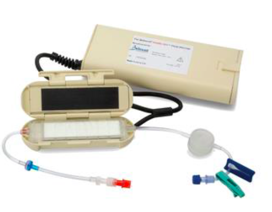
What is in our packs?: Buddy lite fluid warmer, space blankets, EasyWarm Active Self-Warming Blanket

Our second speaker was Dr Andrew Peacock, a part time emergency medicine physician and part time expedition doctor. In the course of his travels around the world he has also developed incredible skill as an award winning photographer. We heard about his recent trip to Denali (Mt McKinley) which, at 6190m above sea level, poses the dual challenge of freezing temperatures and altitude. The main issues Andrew faced on Denali were altitude sickness, frostbite and hypothermia.

http---cdn.cnn.com-cnnnext-dam-assets-171120111021-andrew-peacock-image-4

(Photo credit: CNN, Andrew Peacock)

Altitude related medical issues can occur at altitudes above 2500m but this isn’t the only risk factor; rate of ascent and previous medical issues at altitude are more relevant predictors.

Altitude sickness comprises a spectrum ranging from AMS (Acute Mountain Sickness) through to HAPE (High Altitude Pulmonary Oedema) and HACE (High Altitude Cerebral Oedema).

Symptoms

AMS: headache, nausea, anorexia, dizziness, general malaise

HAPE: dry cough, decreased exercise tolerance, shortness of breath

HACE: headache, nausea, vomiting, truncal ataxia, altered mental status

Treatment

Mild AMS can be treated by stopping the ascent, acetazolamide (125mg bd) and simple analgesia. However both HAPE and HACE (and severe AMS) warrant descent and supplemental oxygen. In addition HACE can be treated with dexamethasone (8mg stat dose then 4mg q6h) and HAPE with nifedipine (30mg slow release bd)

Andrew also provided a broader classification for hypothermia which is easier to apply in the field and is a helpful guide to treatment

Mild: Shivering and normal mental function (temp 32-35 degrees)

Severe: Absence of shivering and abnormal mental function (temp <32 degrees)

Andrew’s top tip for managing frostbite was to avoid rewarming the affected digits until you can guarantee that further frost bite will not occur. Then, rewarm in a bath of 37-39 degrees. This is a painful process and requires liberal analgesia. Finally, splint, elevate and protect.

Andrew’s beautiful travel photographs can be seen on his website   www.footloosefotography.com and on his instagram page https://www.instagram.com/footloosefotography/?hl=en.

If they dont give you an acute case of wanderlust, then nothing will.

References and resources

  1. https://lifeinthefastlane.com/ccc/hypothermia/
  2. https://coreem.net/core/acute-mountain-sickness/
  3. CNN – The expedition doctor who photographs his extreme travels
  4. Wilderness Medicine Society
  5. expeditionmedicine.com.au
Posted in General PH&RM | Tagged | Leave a comment

Education Day – April 4th 2018

Slide1

Posted in General PH&RM | Leave a comment

Blue Sky Trauma from Umbara Base Hospital Part Three

Decision Nodes in trauma care

An algorithm is an illustration of a series of medical decisions that address certain patient specific conditions outlining appropriate responses intended to lead to an outcome.

The goals of an algorithm are to identify diagnostic alternatives, treatment options, and outcomes by weighing decision benefits against risks and costs.

These algorithms can be explicit and published based on best evidence such as in Fig. 1 or individual critical decision making based on expertise and experience.

Decision pathways reach knots or nodes, from which several options are available.

A primary benefit of a well – developed algorithm is that it focuses the reader on the critical decision points in any clinical scenario and specifically lists the input data that leads to a decision. Algorithms can be applied to specific problems, processes, or diseases. They allow for evolution of a disease related topic with new information or clinical conditions that may affect decision making later in the time course of a situation. They can convey the scope of a clinical condition from presentation, through testing and assessment, followed by a clinical judgment and action leading to an eventual outcome. Annotations are added to appropriate points on the algorithm and are necessary for all decision nodes. The purpose of the annotation is to explain all critical factors affecting decisions in as concise a manner as possible. The primary advantage of an algorithm is that it can summarize an evidence based guideline into an easily and quickly available practice protocol for use in the clinical care setting. Fig. 1 is an example of such an algorithm published by the Western Trauma Association.

WTA Pelvic fracture management algorithm.

Gun Shot Wounds (GSW) to abdomen

In countries such as Australia which have very low volume of GSWs to the abdomen, there is a lower threshold for explorative laparotomy versus selective conservatism. The latter may involve observation for peritonism/bleeding and/or performing CT to look for tangential injury without peritoneal violation as is done in high penetrating GSW volume North American trauma centres. CT can be used to ascertain bullet trajectory and can assist in detecting retroperitoneal injury.

In general, management of GSWs such be based on objective physical examination, findings and diagnostic investigations and not on entry and exit sites or sizes.

To retrieve the bullet, or not to retrieve in Operating Theatre?

Retrieved bullet

There are only a few indications to retrieve the bullet. These include bullets found in joints, CSF, or the globe of the eye. Fragments leading to impingement on a nerve or a nerve root, and bullets lying within the lumen of a vessel, resulting in a risk of ischemia or embolization, should be removed. Rare indications are lead poisoning caused by a fragment, and removal that is required for a medico-legal examination. In all other cases the indication should be critically reviewed.

Prehospital blood transfusion in trauma

Many prehospital teams carry refrigerated blood. This can be warmed with the Buddy Lite (Fig. 3). The Buddy Lite can heat 4.4 liters of fluid or blood to 38°C on a single charge. Its major limitation is that it is unable unable to heat fluid that is given rapidly.

Coagulation tests in Major trauma

As well as ordering INR/aPTT all major trauma patients suspected of bleeding should have fibrinogen level sent. Hyperfibrinolysis in major trauma (detected by fibrinogen level/viscoelastic tests) is associated with worse outcomes. It is treated with cryoprecipitate transfusion.

Belmont buddy Lite fluid warmer

Code Crimson

‘Code Crimson’ is a term that has commonly been used by hospital-based teams managing patients with life-threatening haemorrhage that is refractory to resuscitation. The purpose of a ‘Code Crimson’ activation is to streamline patient access to definitive intervention, including an operating theatre or interventional radiology suite. This has recently been formalised in NSW and can be activated by prehospital teams.

Traumatic Brain Injury

Patients who fail aWPTAS with brain injury and who do not have a surgically evacuable intra-cranial lesion are at high risk of suboptimal management. Amnesia is only one symptom and symptoms may also include fatigue, dizziness, headache, cognitive and psychosocial changes. These patients may acutely require referral for in-patient Brain Injury Unit management prior to rehabilitation.

Posted in Cases, Trauma | Tagged , , , | Leave a comment

Clinical Governance Day Weds 21st March

Posted in General PH&RM | Leave a comment

Heat Stress

With Sydney temperatures consistently reaching the high 30’s and early 40’s recently it is perhaps timely to revise heat related illness. It is also worth considering the steps we can take to keep ourselves cool in the same environmental conditions.

Heat related illness covers a spectrum which, at the most extreme end, includes heat stroke. This is defined as hyperthermia >40 degrees, associated with neurological dysfunction. The neurological dysfunction can manifest in many ways including ataxia, delirium, seizures and coma. The patient may also be tachycardic and hypotensive (echo may show either a hypodynamic or hyperdynamic LV). Sweating may be absent. Unrecognised heat stroke carries a mortality of up to 30%.

Management

Aggressive cooling in the field can prevent a cascade of organ dysfunction and significantly improve prognosis. Initial management and cooling methods will be dictated by resources and environment but consider:

  • Moving the patient into the shade
  • Using pack equipment to create some shade e.g. the warming blanket could be used as a canopy if bystanders are able to hold it aloft.
  • Remove the patients clothing
  • Place wet towels or sheets over the patient (road crew may have some)
  • Place ice packs in axillae, groin, neck
  • Try to create some airflow over the patient if possible (not always practicable in the prehospital environment)
  • Stop cooling at approximately 38-38.5 degrees core temp to avoid overshoot hypothermia.
  • Paracetomol is ineffective

Don’t forget standard resuscitation measures:

A = RSI if patient is obtunded and unable to protect airway

B = Ventilate as required. Be aware that pulmonary oedema and ARDS may accompany heat stroke if you are having difficulty ventilating

C = iv fluids may be needed but don’t equate hyperthermia with hypovolaemia. Depending on clinical circumstances, the patient may be either mildly or profoundly volume depleted. Be aware of dilutional hyponatraemia in endurance athletes consuming lots of sports drinks. This can be an important differential for altered LOC and seizures.

Inotropes may be required if shock is refractory to fluid resuscitation but be aware that adrenaline and noradrenaline will cause vasoconstriction that may impair cooling.

D = Benzodiazepines for seizures, don’t forget to check a BSL

Self Care

Self-care is important on these missions as crew members are being exposed to the same environmental conditions as the patient and may have had to exert themselves to get there (winch, bush walk etc.) Make sure you are well hydrated pre mission and carry additional water. Urine colour is the best guide to your hydration status. Ice packs are available in the ready room and can be used by crew if needed. Consider your uniform sunhat if your head is exposed. Sun cream is also available in the ready room. If you are feeling unwell due to heat, communicate this immediately to your crew.

Heat stress

Posted in General PH&RM | Tagged | Leave a comment

Double Pumping Vasoactive Drugs

A significant number of our critical care patients are dependent on vasoactive drugs, so it is worth reviewing the process for managing these infusions during inter-hospital transfers.

pastedgraphic-2

The priming volume of the distal lumen of a central line is 0.44mls. Therefore an infusion running at 10mls / hour could take approximately 3 minutes to reach the patient if started de novo. The half-life of inotropes such as adrenaline and noradrenaline is short, approximately 1 minute. This leaves the patient vulnerable to a period of hypotension. Corrective bolusing of vasoactive drugs is not recommended as it can lead to large, and potentially detrimental, swings in blood pressure. Therefore a smooth transition between syringe pumps is recommended to maintain haemodynamic stability.

There are a number of different methods for double pumping but one suggested protocol is as follows:

  1. Leave infusion number 1 running at its current rate (100%)
  2. Commence infusion number 2 at 50% of the rate of infusion number 1
  3. Wait for a small kick in blood pressure to indicate that the second infusion is reaching the patient. This can be up to but should not exceed SBP 20mmHg
  4. Immediately increase infusion number 2 to 100% (the current rate of infusion 1) and reduce infusion number 1 back to 50%.
  5. Reduce infusion 1 back to zero incrementally over a few minutes ensuring that BP doesn’t drop.

Tips and tricks

  1. Avoid the use of a 3-way tap if you have enough ports to manage without. Three-way taps increase the complexity of the process and the likelihood of an error. No matter how intelligent you think you are, 3-way taps have a habit of embarrassing you. If you really must use a 3-way tap make sure both taps are “on” to the patient during the process and then make sure the correct tap is left “on” at the end.
  2. Dedicate a member of the HEMS team to the double pumping process and avoid being interrupted during it.
  3. Use a dedicated line for vasoactive drugs to prevent inadvertent bolusing
  4. Label the line and the syringe driver clearly to prevent confusing the vasoactive drug with sedation (which can be, and often is, bolused)
  5. If you are about to embark on a long transfer you may want to replace a single strength vasoactive drug with double strength. Take this into account when double pumping. If infusion 2 is double strength it will need to be started at 25% of infusion 1 and then increased to 50% once the BP kick is seen.
  6. Be patient. Depending on the infusion rate and the dead space in the catheter lumen, the process can take several minutes. It is worth taking the time to do it smoothly rather than trying to speed it up by increasing infusion rates or bolusing.

Further reading:

Practical considerations in the administration of i ntravenous vasoactive drugs in the critical care set ting the double pumping or piggyback technique–part oneIntensive and Critical C

An in vitro evaluation of infusion methods using a syringe pump to improve noradrenaline administrationActa Anaesthesiologica Scandinavica 2014 GENAY

Practical considerations in the administration of intravenous vasoactive drugs in the critical care settingIntensive and Critical Care Nursing 2004 Morrice

Management of the changeover of inotrope infusions in childrenIntensive and Critical Care Nursing 2004 ArinoChangeovers of vasoactive drug infusion pumps impact of a quality improvement programCritical Care 2007 Argaud

Posted in General PH&RM | Tagged | 1 Comment